Figure 1.
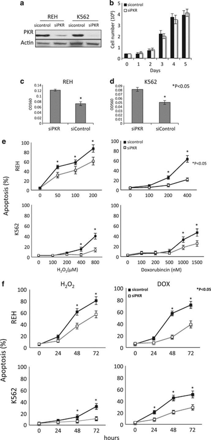
Loss of PKR inhibits apoptosis and promotes survival in response to cellular stress. (a) REH and K562 cells were transduced with lentivirus expressing either PKR SiRNA (SiPKR) or scrambled control SiRNA (Sicontrol). Knockdown of PKR expression was confirmed by western blot. (b) 0.4 × 10⁶ of both Sicontrol and SiPKR cells were cultured for 5 days and cell numbers were counted daily. (c, d) REH or K562 cells were subjected to transwell assay. Invasion across basement membrane was analyzed. (e) Cells were exposed to various concentrations of H2O2 or DOX for 48 h and apoptosis evaluated by flow cytometry using annexin V-PE and 7-AAD staining. (f) REH cells were treated with either 100 μM H2O2 or 400 nM DOX, and K562 cells were exposed to 400 μM H2O2 or 1500 nM DOX. Apoptosis assays were performed at designated time points (0, 24, 48 and 72 h) by flow cytometry. All results are representatives of three independent experiments. *Indicates P<0.05.

