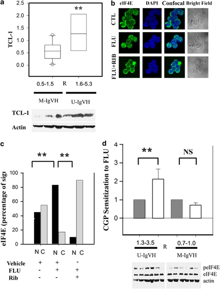
Figure 2.
Ribavirin-mediated sensitization is associated with TCl-1 expression and abrogation of FLU-induced eIF4E nuclear localization. (a) TCL-1 expression is significantly higher in primary CLL samples showing better Ribavirin-mediated sensitization to FLU. TCL-1 expression was assessed in protein extracts of six available samples by western blot. TCL-1 expression was compared using the Mann–Whitney U-statistics (P<0.001). (b) Primary CLL lymphocytes were treated with vehicle (CTL) or FLU IC50 in combination with vehicle (FLU) or 10 μM Ribavirin (FLU+Rib) for 24 h followed by eIF4E staining (green). Nuclear counterstaining was performed with 4',6-diamidino-2-phenylindole (DAPI, blue). Subcellular localization of eIF4E was imaged using a confocal microscope. Bright-field images of the CLL lymphocytes analyzed are shown in the right panel. (c) Nuclear (N, black bars) or cytoplasmic (C, gray bars) eIF4E localization after treatment with vehicle, FLU IC50 or FLU IC50 plus 10 μM Ribavirin is represented as percentage of the fluorescent signal calculated from 15 to 25 cells per condition. The Fisher test indicates that there is a significant difference between the eIF4E nuclear staining patterns after the treatments (**P<0.01, Fisher test). (d) The bars represent the mean sensitization value of CGP57380 on FLU sensitivity in seven M-IgVH and seven U-IgVH samples (open bars) with respect to paired vehicle-treated samples (gray bars) (y axis). eIF4E phosphorylation is shown in representative samples from each group.

