Fig. 9.
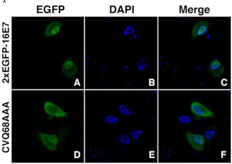
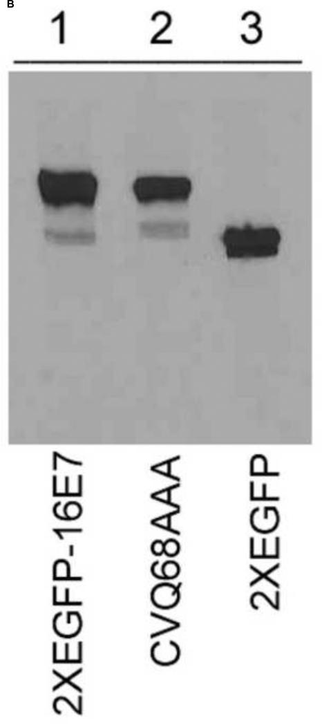
A. Analysis of the effect of CVQ68AAA mutation on the nuclear localization of 2xEGFP-16E7. HeLa cells were transfected with 2xEGFP-16E7 (panels A, B and C) or 2xEGFP-16E7CVQ68AAA (panels D, E and F) plasmids, and the localization of the expressed proteins was analyzed by confocal fluorescence microscopy. Panels A and D represent the fluorescence of the EGFP, panels B and E the DAPI staining of the nuclei, and panels C and F the merge. B. The 2xEGFP-16E7CVQ68AAA mutant is expressed properly in HeLa cells. HeLa cells were transfected with 2xEGFP-16E7 (lane 1), 2xEGFP-16E7CVQ68AAA (lane 2) and 2xEGFP (lane 3). Cell lysates were prepared 24 h post transfection and probed with a GFP antibody.


