
Figure 4.
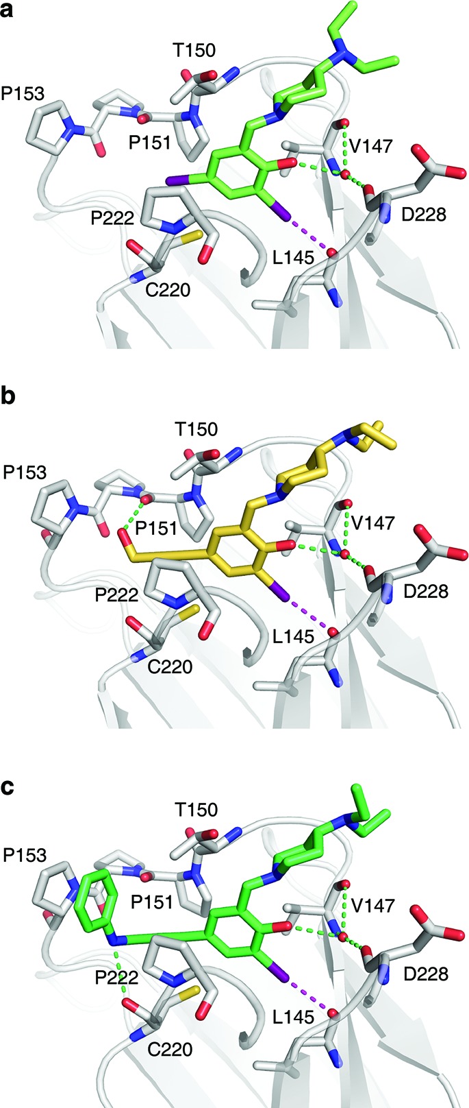
Crystal structures of Y220C–ligand complexes. Shown are the binding modes of 4 (a), PhiKan5116 (b), and PhiKan5196 (c). The protein is shown as a gray cartoon representation with selected residues highlighted as stick models. The halogen bond between the iodine and the carbonyl oxygen of Leu145 is indicated by a broken magenta line; additional polar interactions with the protein are shown as green broken lines.