
Figure 2.
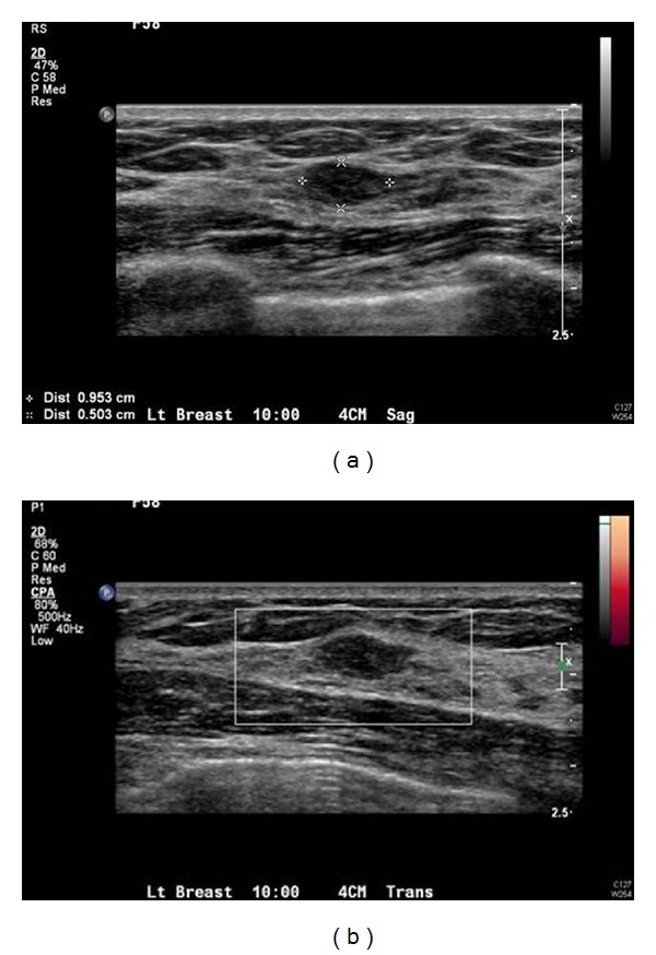
(a) Ultrasound evaluation in the upper inner breast demonstrates an oval, hypoechoic lesion, corresponding to the lesion seen on MRI (though slightly larger in dimension) with minimally irregular margins. (b) No increased vascularity.
Official websites use .gov
A
.gov website belongs to an official
government organization in the United States.
Secure .gov websites use HTTPS
A lock (
) or https:// means you've safely
connected to the .gov website. Share sensitive
information only on official, secure websites.
(a) Ultrasound evaluation in the upper inner breast demonstrates an oval, hypoechoic lesion, corresponding to the lesion seen on MRI (though slightly larger in dimension) with minimally irregular margins. (b) No increased vascularity.