
Figure 6.
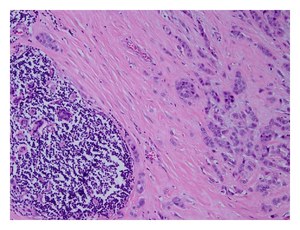
Infiltrating carcinoma cells, seen on the right side of the image, are present adjacent to an atypical lymphoid infiltrate consistent with chronic lymphocytic leukemia, present on the left side of the image (mastectomy specimen, hematoxylin & eosin stain, 100x magnification).