
Figure 1.
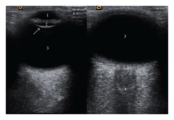
Transverse gray-scale US image that shows normal anatomy of ocular structures: anterior chamber (1), lens (2), vitreous body (3), in the conal region, the hypoechoic central band (4) corresponding to optic nerve sheath complex, and the retrobulbar fat (5).