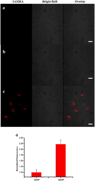
Figure 4.
Switchable aptamer micelle flares respond to target molecules in HeLa cells. Confocal microscopy fluorescence imaging of intracellular ATP molecules with 1 μM (a) aptamer switch probe without diacyllipid conjugation, (b) CSMFs and (c) SAMFs. Left panels are TAMRA fluorescence pseudo-colored red, middle panels are the bright-field image and right panels are the overlay of TAMRA fluorescence and the bright-field image. Scale bar: 50 μm. (d) Cell-associated fluorescence (TAMRA) of cell populations treated with 1 μM CSMFs and SAMFs, as determined by flow cytometry.

