
Figure 5. TGFα activates EGFR through a distinct orientation of JM helices.
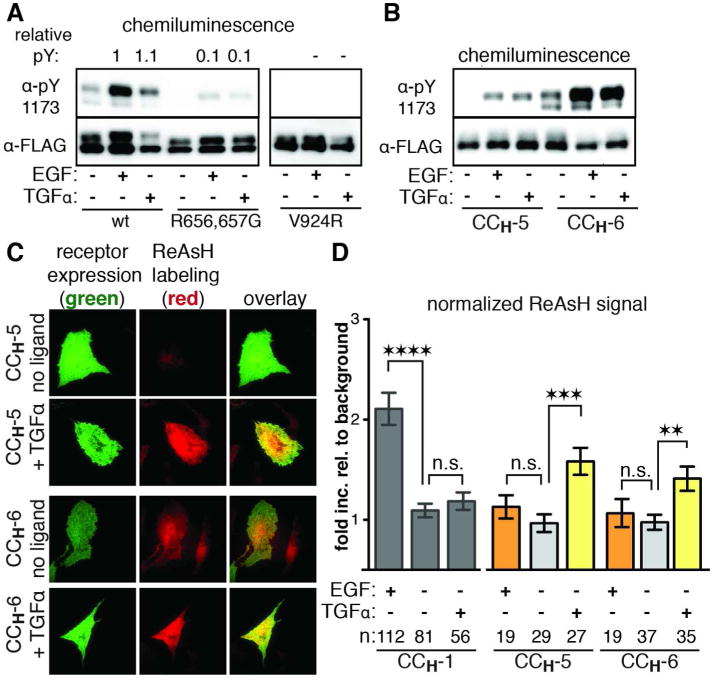
(a) Western blot analysis of wt, R656,657G, and V924R EGFR stimulated with EGF or TGFα. See also Figure 3A. (b) Western blot analysis of CCH-5 and CCH-6 EGFR stimulated with EGF or TGFα. (c) Representative TIRFM images of cells expressing CCH-5 and CCH-6 EGFR that were labeled with ReAsH in the presence or absence of TGFα. See also Supporting Figure S4. (d) Quantification of TIRFM results as a fold increase relative to background that is normalized for receptor expression levels. n is the number of cells quantified. Error bars represent the standard error. ** p<0.01, *** p<0.001, **** p<0.0001, one-way ANOVA with Bonferroni post-analysis accounting for multiple comparisons.
TGFα leads to a structural transition in the JM helices, allowing for CCH-5 and CCH-6 to be labeled with ReAsH. These findings suggest that activation of EGFR by TGFα occurs through a JM helical orientation that is distinct from the antiparallel coiled coil determined for activation by EGF.