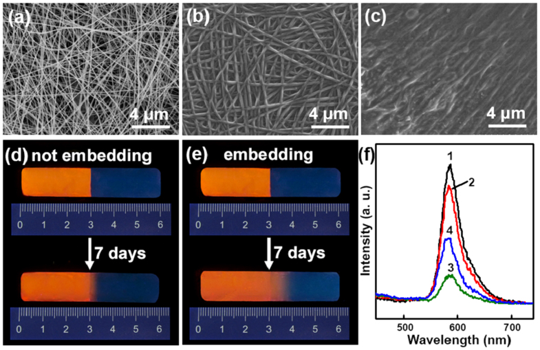
Figure 4. Morphology and molecular diffusion process in healed hydrogels embedded with and without the healing layer.
(a–c) FESEM images of (a) nanofibers without redox initiators, and the healing layer (b) as-prepared and (c) after embedding into bulk hydrogels. (d,e) Fluorescent images and (f) fluorescence spectra of hydrogels constructed by connecting together two hydrogel blocks with and without 0.05 wt% rhodamine B respectively stored for 7 days: (d) without and (e) with the healing layer embedded between the two merged hydrogel blocks. (f) The corresponding fluorescence spectra of hydrogels after stored for 7 days: at position of (1,2) 2.8 cm and (3,4) 3.2 cm for (1,3) sample (d) and (2,4) sample (e).

