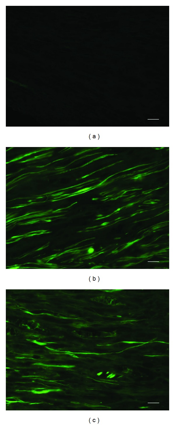
Figure 3.

GAP-43 immunofluorescence staining was observed at 9 mm proximate to the crushed site in the control, Epimedium, and icariin groups at week 1 after the operation. (a) GAP-43 staining was hardly seen in the control group. (b) Evident GAP-43 staining was observed in the Epimedium extract group. (c) In the icariin group, GAP-43 staining was detectable but the density was lower than the Epimedium extract group. Scale bar: 5 μm.
