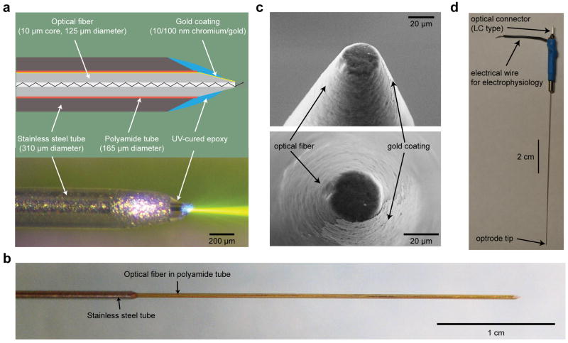
Figure 1. The structure of the coaxial optrode.
(a) Cross sectional schematic (top) and side view photograph (bottom) of the coaxial optrode showing main constituent parts. The center optical fiber has a 10 μm diameter optical aperture (exposed core of the fiber) leading to highly directional light output as visualized in dye-doped saline. The saline contains ~1 μM fluorescein with fluorescence excited by 473 nm laser light (bottom). (b) A larger scale image of the device where the reinforcing thin stainless steel jacket (310 μm outer diameter) was kept few centimeters away from the tip leading to a thinner tissue penetrating portion of the shaft (diameter 165 μm). (c) Scanning electron microscope images of the coaxial optrode tip. The tip was polished to a flat circular tip of diameter 20-30 μm to separate the optical aperture from the surrounding gold recording electrode layer. This design reduced light-induced artifacts in the electrophysiological recordings to noise level. (d) Full length photograph of the coaxial optrode showing its electrical and optical connections at the distal end.

