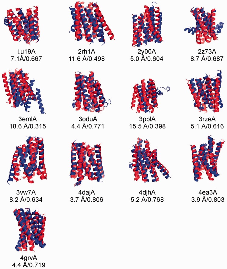
Fig. 3.
Superposition of the first model (blue) and the X-ray structure (red) in the transmembrane regions for 13 known GPCRs. Models are generated by I-TASSER with contact restraints from MemBrain. All GPCR templates and the homologous templates with a sequence identity >30% detectable by PSI-BLAST have been excluded during I-TASSER simulations

