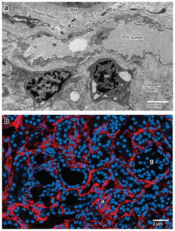
Figure 1.
(a) Electron microscope image of myofibroblasts (MFs) in the interstitial space of a kidney from a patient with chronic kidney disease. Note the abundance of rough endoplasmic reticulum in these cells due to high ribosomal activity, and note the markedly expanded interstitial space with collagen fibers.
Abbreviations: CBM, capillary basement membrane; EC, endothelial cell; PTC, peritubular capillary; TBM, tubule basement membrane. (b) Confocal image of α–smooth muscle actin–expressing MFs (red ) in adult diseased mouse kidney. Abbreviations: a, arteriole; g, glomerulus.

