Figure 4.
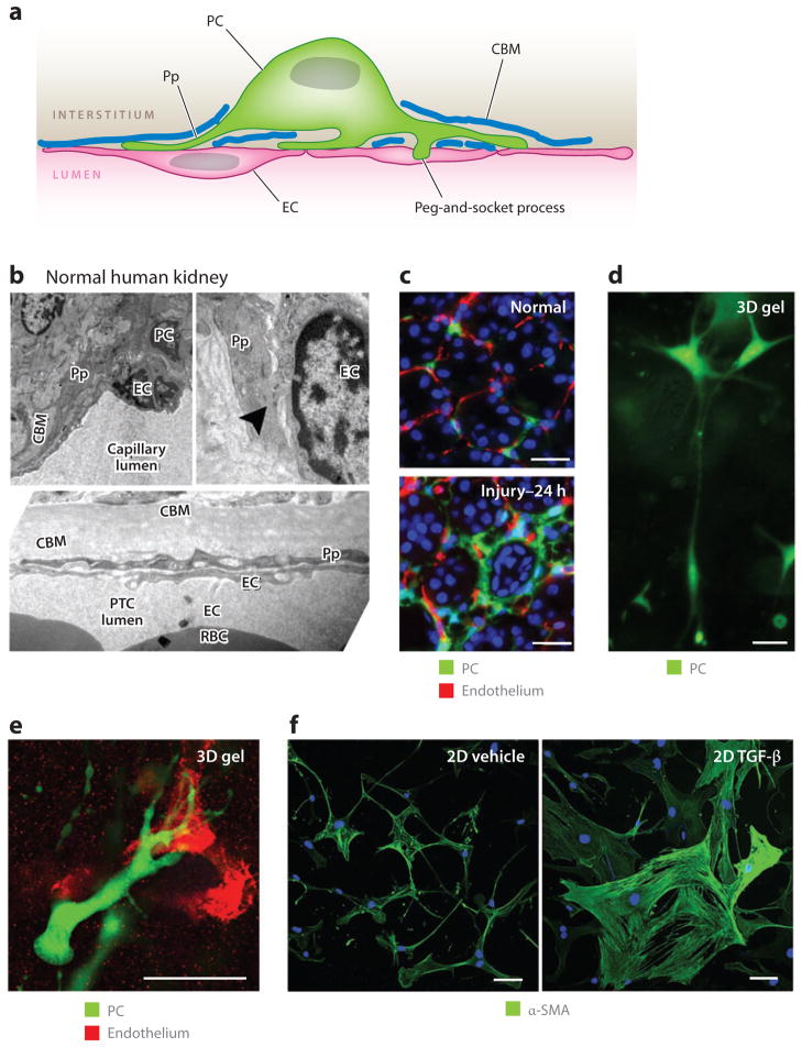
Pericytes (PCs) in the kidney: definitions, functions, and response to cytokines. (a) A PC attached to an endothelial cell (EC) and partially embedded in a duplication of the capillary basement membrane. Note the specific attachment of PC processes (Pp) and the cell body to the EC at several different sites. (b) Electron microscope images of normal human kidney cortex. Shown are peritubular capillaries (PTCs), in which ECs and PCs or Pp are visible. The tubule basement membrane is clearly visible. Note the peg-and-socket process (arrowhead ) at the upper right. (c) PCs attach to ECs in normal mouse kidney, but only 24 h (prior to mitosis) after the onset of obstructive injury, the PCs detach, spread, and migrate from the ECs. (d) PCs in three-dimensional (3D) collagen gel that exhibit long cytoplasmic processes that extend the length of more than 10 cell bodies. (e) PCs in 3D collagen gel home to and bind by attachment specifically to capillary tubes composed of endothelial cells. (f) Kidney PCs cultured on gelatin matrix, stained for α–smooth muscle actin (α-SMA). Note that, in control conditions, PCs show weak α-SMA expression and many long fine processes and elongations, but 24 h after exposure to transforming growth factor (TGF)-β, the PCs change shape, spread, lose their long cytoplasmic processes, upregulate α-SMA, and show distinct cytoplasmic filaments. Scale bars, 25 μm. Abbreviations: 2D, two-dimensional; CBM, capillary basement membrane; RBC, red blood cell.

