
Figure 2. Morphological characterization of fixed BMSC transfer onto PDMS under SEM.
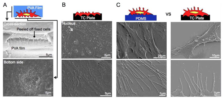
(A) A cross-sectional image of stripped Cell-PVA film indicates complete integration of fixed stromal and PVA film without any gap. This image shows a cross-section of layered stromal cells. The bottom side Cell-PVA film shows submicron scale rough surface as a result of torn intracellular components. (B) A tissue culture plate after being stripped off a cell-PVA film shows intracellular components of remaining cellular mass that formed firm attachment on substrate. (C) Morphological comparison of fixed-PDMS transferred BMSCs and fixed BMSCs shows integrally transferred fixed BMSCs surface on PDMS membrane even in nanoscale resolution.