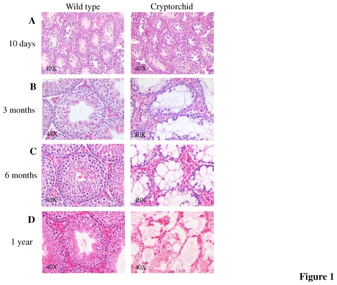
Figure 1. Progressive degeneration of cryptorchid testis.
(A) Male cryptorchid testis shows no differences at day 10 compared to wild-type control. (B) By 3 months fewer differentiating germ cells are present in cryptorchid testes with big Sertoli cell intracellular vacuoles and dilated intercellular spaces. The germ cells are almost all absent by 6 months (C). (D) One year old cryptorchid testis showed severely disrupted tubular structure with single germ cells.

