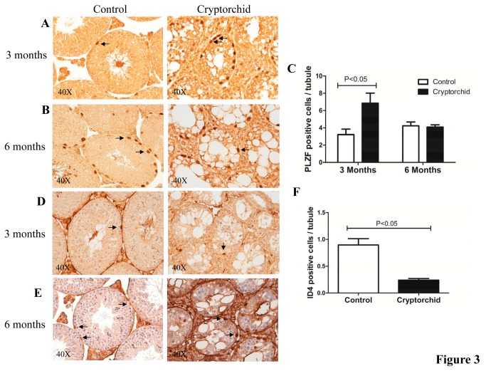
Figure 3. Immunohistochemistry (IHC) analysis for PLZF and ID4 staining in cryptorchid testes.
Cross sections of cryptorchid and control testes stained with antibody for PLZF at 3 months (A) and at 6 months (B). Quantitative comparison of PLZF positive cells (arrows) per seminiferous tubule between cryptorchid and control mice at 3 months and 6 months (C). Cross sections of cryptorchid and control testes stained with antibody for ID4 at 3 months (D) and at 6 months (E). Quantitative comparison of ID4 positive cells (arrows) per tubule between cryptorchid and control mice at 3 months and 6 months (F). Data represent the mean ± SEM.

