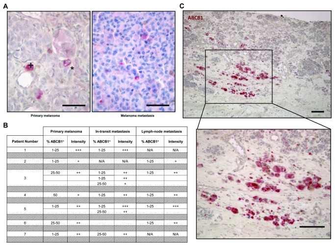
Figure 6. ABCB1 expression in primary human melanomas and corresponding metastases.
A) Representative examples of primary melanoma (left) and melanoma metastasis (right) immunostained for ABCB1 (scale bar, 150µm; +, cytoplasmic staining; *, membranous staining).
B) Summary of semi-quantitative analysis, showing estimated percentage of ABCB1+ cells and signal intensity (no staining = 0, weak signal = +, moderate signal = ++, strong signal = +++; N/A, not applicable).
C) Primary melanoma immunostained for ABCB1 (top) and higher magnification of the boxed area (bottom) (scale bar, 300µm).

