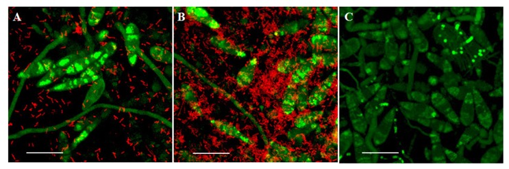
Figure 1. Confocal images of the interaction assay of M. oryzae and L. enzymogenes wild-type strain C3.

M. oryzae expressing a green fluorescent protein and L. enzymogenes expressing a dsRed fluorescent protein at 3hpi (A) and 9 hpi (B), and a mock inoculated sample (C). The long, thin structures are hyphae, whereas the tear-drop shaped structures are conidia. The smaller red rod shapes are bacteria. M. oryzae conidium size ranges from 20 to 30 µm. Scale bar: 20µm.
