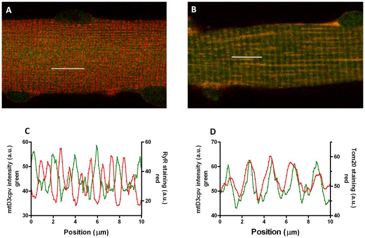
Figure 1. Localization of 4mtD3cpv in FDB muscle fibers.
A and C: mitochondrial cameleon (4mtD3cpv) fluorescence (green) and anti-RyR antibody staining (red) in a merged image (A) and intensity profiles (C). B and D: mitochondrial cameleon (4mtD3cpv) fluorescence (green) and anti-Tom20 antibody staining (red) in a merged image (B) and intensity profiles (D). Segments 10 μm long were scanned on the images as indicated by the white bars.

