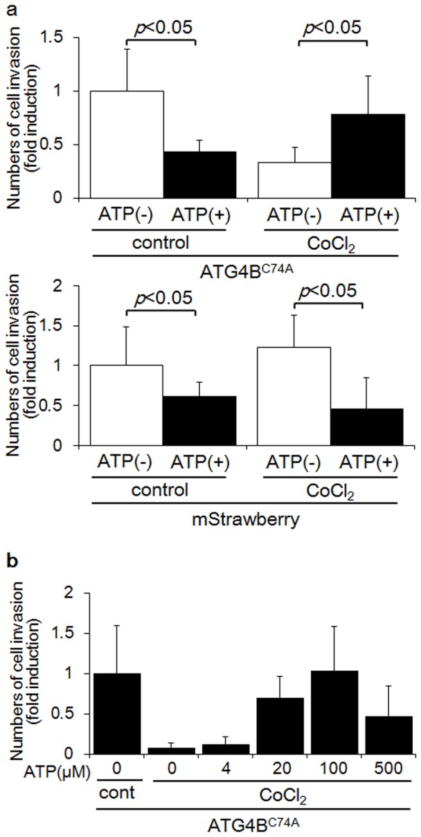
Figure 4. ATP supplementation recovered the suppression of invasion in autophagy-deficient HTR8/SVneo cells.

a) Invasion assays were performed with HTR8-Atg4BC74A mutant cells (upper panel), the autophagy-deficient EVT cell line, or HTR8-mStrawberry cells (lower panel), the control cell line, under DMSO (control) or 250 µM CoCl2 in the presence (black bars) or absence (white bars) of 100 µM ATP for 48 h. The Y-axis indicates the number of invading cells. b) Invasion assays were performed with HTR8-Atg4BC74A mutant cells under 250 µM CoCl2 in the presence of increasing concentrations of ATP, as indicated for 48 h. The Y-axis indicates the number of invading cells. These experiments were independently performed at least three times.
