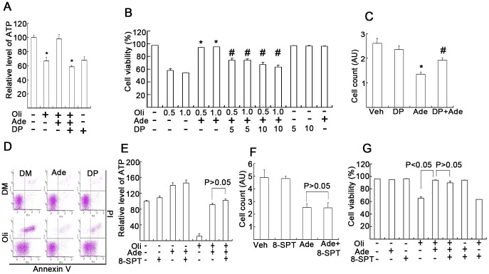
Figure 4. Ade transportation, but not Ade receptors, is required for Ade to exert its cytotoxic or cytoprotective effects.
(A) K562 cells cultured in glucose-free culture medium were treated with Oli (0.5 µg/ml), Ade (2 mM) and DP (10 µM) for 3 h, followed by HPLC ATP assay. Mean ±SD (n = 3). *p<0.05 vs. Oli+Ade treatment. (B) K562 cells were cultured and treated in the d-glucose-free medium as indicated for 18 h, and cells were then stained with Annexin V/PI followed by flow cytometry. Viable cells are shown. Mean ±SD (n = 3). *p<0.05 each compared with Oli treatment alone; #p<0.05 each compared with Oli+Ade combination treatment. (C) K562 cells were cultured in normal medium and exposed to 2 mM Ade and DP (10 µM) for 18 h, cell numbers were counted using a cell counter. Mean ±SD (n = 3). *p<0.05 compared with vehicle control; #p<0.05 compared with Ade treatment alone. (D) As treated in (B), typical flow images are shown (Ade: 2 mM, Oli: 1.0 µg/ml, DP: 10 µM). (E) K562 cells were cultured in d-glucose-free medium and treated with the agents as indicated (Oli: 1.0 µg/ml, Ade: 2 mM, 8-SPT: 10 µM) for 4 h followed by ATP assay. Mean ±SD (n = 3). (F) K562 cells were cultured in normal glucose-containing medium and treated as indicated (Ade: 2 mM, 8-SPT: 10 µM) for 18 h, cell numbers were counted and summarized. Mean ±SD (n = 3). (G) K562 cells were cultured in glucose-free medium and treated as indicated (Oli: 1.0 µg/ml, 8-SPT: 10 µM) for 15 h, cell death was detected by flow cytometry. Mean ±SD (n = 3).

