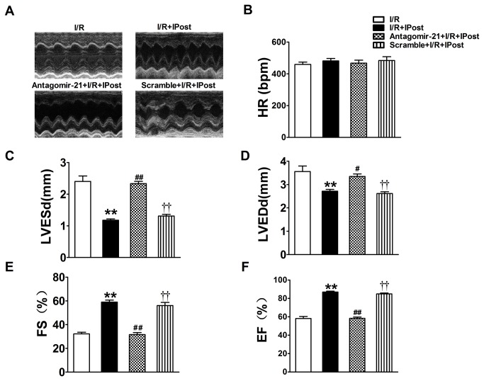
Figure 4. Echocardiographic assessment of left ventricular dimensions and function.
Echocardiography was performed as described in Methods section (A) Representative M-mode echocardiographs from the four groups of mice with different treatments. (B) IPost treatment greatly improved left ventricular function after two weeks of reperfusion. But antagomir-21 pre-treatment exhibited significantly depressed cardiac function recovery during in vivo I/R mouse model, HR: heart rate; LVEDd: left ventricular end-diastolic dimensions; LVESd: left ventricular end-systolic dimensions; EF: ejection fraction; FS: percentage fractional shortening. Data are expressed as mean±SEM, n=5; ** P<0.01 compared with I/R; # P<0.05, # # P<0.01 compared with IPost+I/R group; † † P<0.01 compared with Antagomir-21+IPost+ I/R group.

