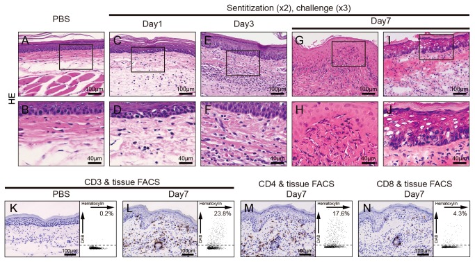
Figure 2. Immunohistochemical analyses of footpads from Pd-induced allergic mice.
Histopathology and immunohistochemical (IHC) analyses were undertaken to identify CD3+, CD4+ and CD8+ cells in footpad tissues. Frozen sections of the footpad tissues were prepared from saline-injected mice or Pd-induced allergy mice (two sensitizations–three challenges) at 7 days after the last challenge. Sections were stained with H&E (C–J), anti-mouse CD3ε (K and L), anti-CD4 (M) and anti-CD8 mAbs (N) and visualized by DAB. Representative photomicrographs are shown. Liquefaction degeneration of basal epithelial layer and infiltration of inflammatory cells into the epithelial basal layer and dermis were observed in Pd-induced allergy mice (G-J), but not control mice (A and B). For IHC analyses, epidermotropism of CD3+ cells was present in the epithelial basal layer and dermis of the Pd-induce allergy mice (L), but not control mice (K). CD4+ T cells were abundant (M) while a small number of CD8+ T cells existed only around blood vessels (N). Representative IHC images were analyzed by HistoQuest software and the data was shown as scatter grams on the right.

