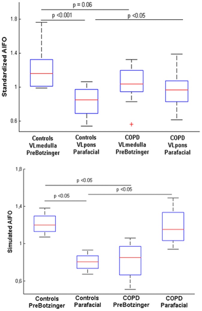
Figure 3. fMRI results of the brainstem respiratory centers at rest.

Top. Amplitude of the low frequency oscillations (AlFO) of the resting state BOLD signal computed in controls and COPD patients. In healthy subjects the AlFO of the rostral ventro-lateral (VL) medulla that contains the pre-Bötzinger complex is higher than the VL medulla of the patients. Conversely, the ALFO of the caudal (VL) pons, which contains the parafacial respiratory group is higher in patients than the VL pons of healthy subjects. Bottom. Simulated AlFO obtained after hemodynamic convolution of the theoretical neural states. For controls, the chosen network scheme is described in Figure 8B, while for COPD patients, the network scheme is described in Figure 8C. Of note the synchronization regime describe in Figure 8D gave the same results as 8C for the simulated AlFO of the BOLD signal.
