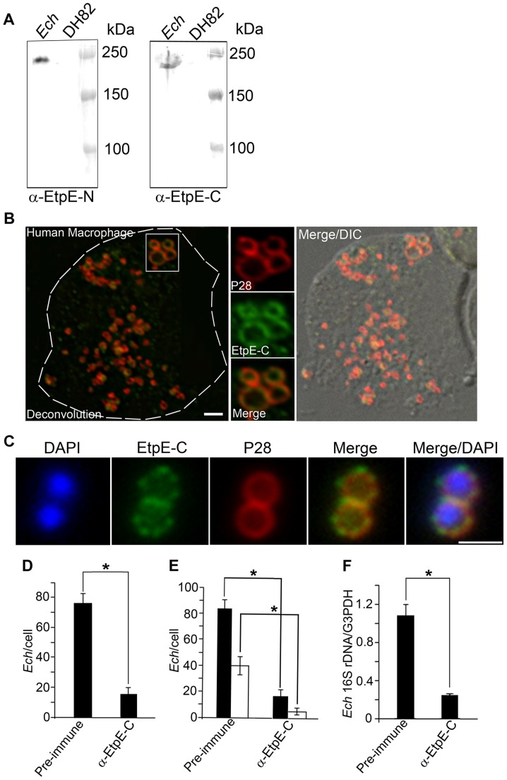
Figure 1. EtpE-C is exposed at the bacterial surface, and anti-EtpE-C neutralizes E. chaffeensis infection in vitro.
(A) Western blot analysis of E. chaffeensis-infected (Ech) and uninfected DH82 cells at 60 h pi using anti-EtpE-N (α-EtpE-N) and anti-EtpE-C (α-EtpE-C). (B) Double immunofluorescence labeling of E. chaffeensis-infected human primary macrophages derived from peripheral blood monocytes at 56 h pi. Cells were fixed with PFA, permeabilized with saponin, and labeled with anti-EtpE-C and anti-E. chaffeensis major outer membrane protein P28. The white dashed line denotes the macrophage contour. The boxed region indicates the area enlarged in the smaller panels to the right. Merge/DIC: Fluorescence images merged with Differential interference contrast image (DIC). A single z-plane (0.4 µm thickness) by deconvolution microscopy was shown. Scale bar, 2 µm. (C) E. chaffeensis was incubated with DH82 cells for 30 min and double immunofluorescence labeling was performed using anti-EtpE-C and anti-E. chaffeensis P28 without permeabilization. DAPI was used to label DNA. Scale bar, 1 µm (see also suppl. Fig. S2). (D) Numbers of E. chaffeensis bound to RF/6A cells at 30 min pi. Host cell-free E. chaffeensis was pretreated with anti-EtpE-C or preimmune mouse serum and incubated with RF/6A cells for 30 min. Unbound E. chaffeensis was washed away, cells were fixed with PFA, and E. chaffeensis labeled with anti-P28 without permeabilization. E. chaffeensis in 100 cells were scored. (E) Numbers of E. chaffeensis internalized into RF/6A cells at 2 h pi. E. chaffeensis was pretreated with anti-rEtpE-C or preimmune mouse serum and incubated with RF/6A cells for 2 h. To distinguish intracellular from bound E. chaffeensis, unbound E. chaffeensis was washed away and cells were processed for two rounds of immunostaining with anti-P28; first without permeabilization to detect bound but not internalized E. chaffeensis (AF555–conjugated secondary antibody) and second round with saponin permeabilization to detect total E. chaffeensis, i.e., bound plus internalized (AF488–conjugated secondary antibody). E. chaffeensis in 100 cells was scored. The black bar represents total E. chaffeensis and the white bar represents internalized E. chaffeensis (total minus bound) (see also suppl. Fig. S3). (F) Infection of RF/6A cells with E. chaffeensis at 48 h pi. E. chaffeensis was pretreated with anti-EtpE-C or preimmune mouse serum and used to infect RF/6A cells; cells were harvested at 48 h pi. qPCR for E. chaffeensis 16S rDNA was normalized with G3PDH DNA. Data represent the mean and standard deviation of triplicate samples and are representative of three independent experiments. *Significantly different (P<0.05).

