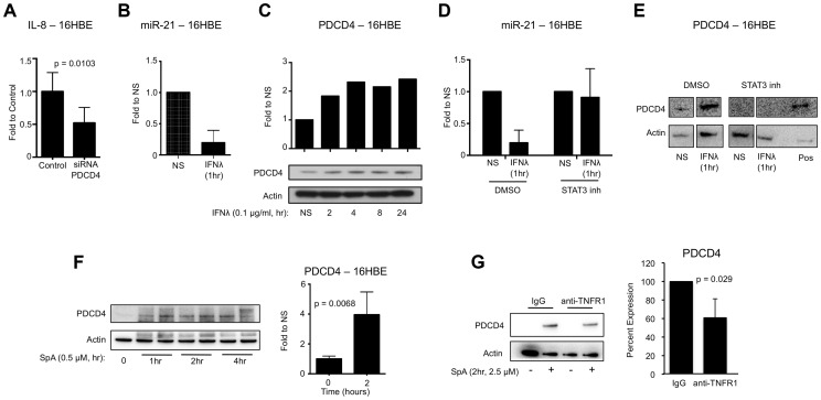
Figure 3. IFNλ regulation of miR-21 and PDCD4.
(A) qRT-PCR analysis of IL-8 in 16HBE cells treated with control or PDCD4 siRNA and infected with USA300, µ ± SD. (B) qRT-PCR analysis of miR-21 in 16HBE cells treated with recombinant IFNλ for 1 hour, µ ± SD. (C) Western blot analysis of PDCD4 in 16HBE cells treated with recombinant IFNλ. (D) qRT-PCR analysis of miR-21 in 16HBE cells pretreated with STAT3 inhibitor or DMSO treated with recombinant IFNλ for 1 hour, µ ± SD. (E) Western blot analysis of PDCD4 in 16HBE cells pretreated with STAT3 inhibitor or DMSO treated with recombinant IFNλ for 1 hour. Lysate from 16HBE cells treated with DMSO and IFNλ was used as a positive control (pos). (F) Western blot analysis of PDCD4 in human epithelial cells (16HBE) following stimulation with S. aureus protein A (SpA), µ ± SD. (G) Western blot analysis of PDCD4 in 16HBE cells treated with anti-TNFR1 or control IgG and SPA for 2 hours, µ ± SD. Data are representative of at least 2 independent experiments.

