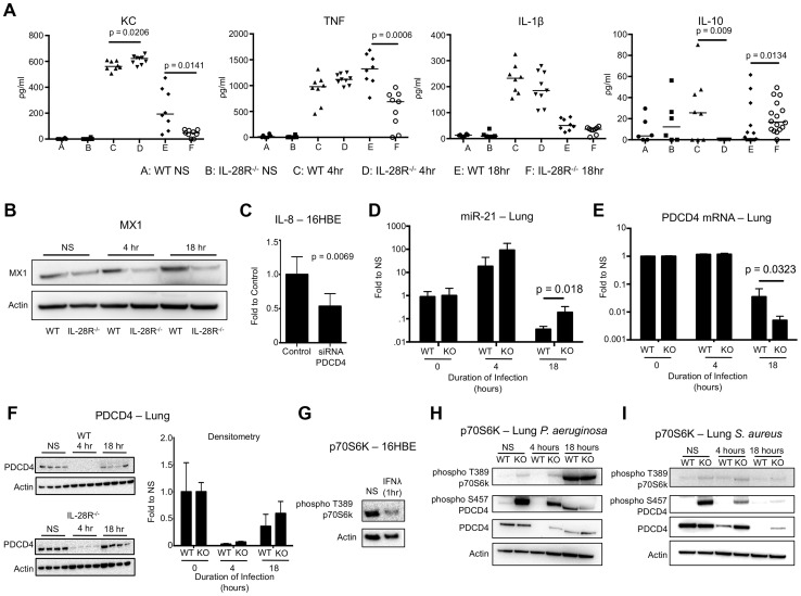
Figure 6. P. aeruginosa induced cytokine, miR-21, and PDCD4 expression in WT and IL-28R−/− mice.
(A) ELISA analysis of individual cytokines in BAL of WT and IL-28R−/− mice. (B) Western blot analysis of MX1 expression in lungs of WT and IL-28R−/− mice following 4 and 18 hours of infection with PAK. (C) qRT-PCR analysis of IL-8 in 16HBE cells treated with control or PDCD4 siRNA and infected with PAK, µ ± SD. (D) qRT-PCR analysis of miR-21 in the lungs of WT and IL-28R−/− mice following infection with PAK, µ ± SD. (E) qRT-PCR analysis of PDCD4 mRNA in the lungs of WT and IL-28R−/− mice following infection with PAK, µ ± SD. (F) Western blot analysis of PDCD4 in the lungs of WT and IL-28R−/− mice following infection with PAK, µ ± SD. (G) Western blot analysis of phosphorylation of p70S6K in 16HBE cells treated with recombinant IFNλ. (H) Western blot analysis of phosphorylation of p70S6K and PDCD4 in the lungs of WT and IL-28R−/− mice following infection with PAK. (I) Western blot analysis of phosphorylation of p70S6K and PDCD4 in the lungs of WT and IL-28R−/− mice following infection with USA300. Data are representative of at least 2 independent experiments.

