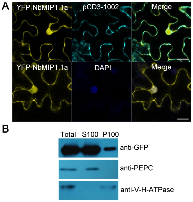
Figure 4. The subcellular localization of NbMIP1.1a in N. benthamiana cells.

(A) Confocal image of the subcellular localization of NbMIP1.1a in leaf epidermal cells. YFP-NbMIP1.1a was transiently expressed in leaves of N. benthamiana via agroinfiltration and imaged at 48 hpi using a Zeiss LSM 710 laser scanning microscope. YFP signal revealed that NbMIP1.1a is present in the cell membrane, cytoplasm and nucleus. PCD3-1002: a CFP-tagged plasma membrane marker [81]. DAPI: staining for nuclei. Scale bar represents 20 µm. (B) YFP-NbMIP1.1a was found in both the soluble fraction and the membrane fraction (upper panel). Protein extracts were centrifuged at 100,000×g to produce crude soluble (S100) and microsomal (P100) fractions. Fractions were analyzed by western blot following separation by SDS-PAGE. The gels were probed using anti-GFP, anti-V-H-ATPase (vacuolar H-ATPase subunit, a vacuolar membrane marker) and anti-PEPC (phosphoenolpyruvate carboxylase, a cytosolic marker) antibodies as indicated.
