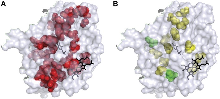
Figure 1.
Positions of the altered CBS amino acid residues on the 3D structure of the truncated CBS protein. The pyridoxine and heme cofactors are shown in black. (A) Forty-four amino acid residues representing 51 of the 58 substitutions are shown in red. Seven of the substitutions were in residues not included in the 3D structure and are not shown. (B) The fourteen residues that, when substituted, resulted in CBS protein variants with altered cofactor sensitivity in vivo. All variants demonstrated altered sensitivity to pyridoxine; the two variants that had altered sensitivity to both heme and pyridoxine (K267E and L345P) are shown in green.

