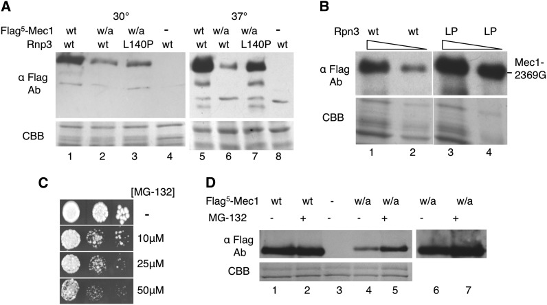
Figure 9.
rpn3-L140P increases the level of C-terminally altered Mec1 derivatives. (A) Mec1-W2368A. Protein extracts were prepared by bead lysis in buffer lacking protease inhibitors from diploid yeast strains CY6172 (MEC1/Flag5-MEC1 RPN3/RPN3), CY6192 (MEC1/Flag5-mec1-W2368A RPN3/RPN3), CY6400 (MEC1/Flag5-mec1-W2368A rpn3-L140P/rpn3-L140P), and BY4743 (MEC1/MEC1 RPN3/RPN3) grown at 30° or 37°; 50 μg of protein was separated by sodium dodecyl sulfate (SDS)-PAGE and the upper portion of the gel was Western-blotted with anti-Flag antibody. The lower part of the gel was stained with Coomassie brilliant blue (CBB). (B) Mec1-2369G. Yeast strains CY6349 (mec1-2369G RPN3 sml1Δ0::KanMX; lanes 1 and 2) and CY6449 (mec1-2369G rpn3-L140P sml1Δ0::KanMX; lanes 3 and 4) were grown in YPD media for 8 hr at 37°; 40 μg (odd lanes) and 20 μg (even lanes) of protein extract was separated by SDS-PAGE and Western-blotted with anti-Flag antibody or stained with CBB. (C) BY4742 was grown in media containing proline as the nitrogen source and 0.003% SDS for 3 hr, then serial dilutions were plated onto identical synthetic complete media with the indicated amount of MG-132. (D) CY6172 (MEC1/Flag5-MEC1; lanes 1 and 2), BY4743 (lane 3), and CY6192 (MEC1/Flag5-mec1-W2368A; lanes 4–7) were grown in media containing proline as the nitrogen source and 0.003% SDS for 3 hr at 37°. MG-132 was added to a final concentration of 75 μM (+) or the equivalent volume of dimethylsulfoxide (−) and the cells were grown for an additional 2 hr. Extracts were prepared by glass bead lysis and 30 μg was separated by SDS-PAGE. The upper portion of the gel was Western-blotted with anti-Flag antibody. The lower portion was stained with CBB. Lanes 6 and 7 are an overexposure of lanes 4 and 5.

