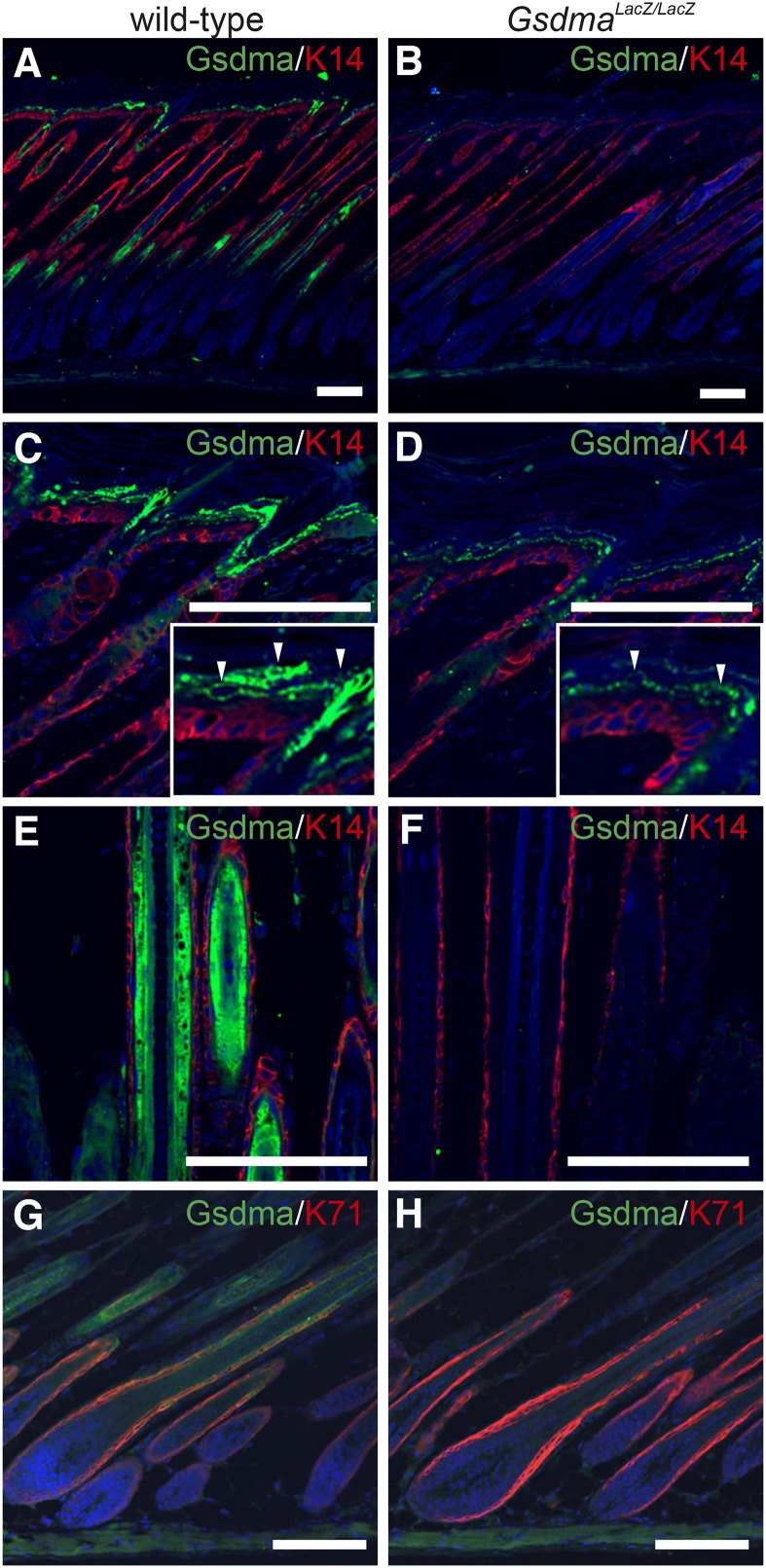
Figure 4.
Distribution of Gsdma and Gsdma3 proteins. Immunohistological detection of Gsdma and Gsdma3 proteins in skin of wild-type (A, C, E, and G) and GsdmaLacZ/LacZ (B, D, F, and H) mice at P8. Magnified images are provided to show Gsdma protein (arrowheads) in the suprabasal cell layer. K14 and K71 were used as a marker for the basal cell layer and IRS, respectively. Scale bars are 25 μm.

