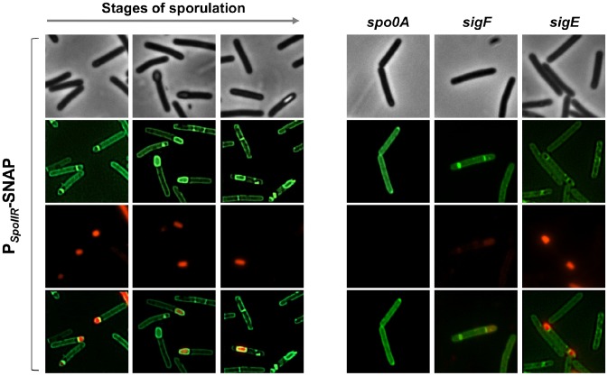
Figure 5. Fluorescence of a PspoIIR-SNAP fusion in strain 630Δerm and in a spo0A, sigF or sigE mutant.
Cells of the C. difficile 630Δerm strain, and of the spo0A, sigF and sigE mutants carrying a PspoIIR-SNAPCd transcriptional fusion in a multicopy plasmid were collected 24 h of following inocculation in SM broth. Cells were labelled with the fluorescent substrate TMR to allow localization of SNAPCd production driven by the spoIIR promoter, stained with the DNA marker DAPI and the membrane dye MTG and examined by phase contrast and fluorescence microscopy.

