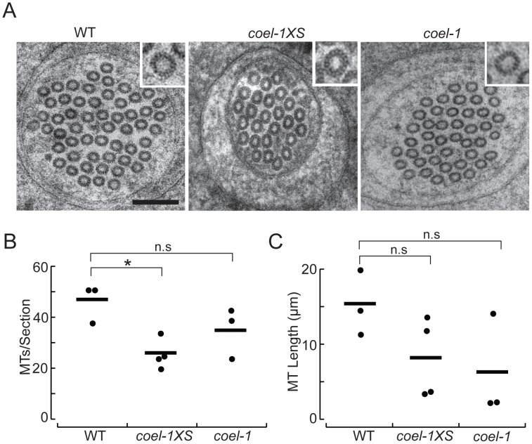
Figure 5. coel-1 activity influences microtubule number and length, but not protofilament count in PLM touch receptor neurons.
A. High-resolution electron micrographs of thin (50 nm) sections of PLM touch receptor neurons in wild-type (WT), coel-1XS and coel-1(tm2136) animals. Insets show a single microtubule profile, revealing the protofilaments. Scale bar, 100 nm. B. Number of MTs per section as a function of genotype. Bars are the mean values and filled circles are the average values in each dataset. C. Microtubule length as a function of genotype. Bars are the mean and filled circles are the number of serial section datasets tested. Microtubule (MT) length computed from: L = 2Na/T, where N = the average number of MTs/section, a = total length of serial reconstruction and T = number of MT endpoints observed [25]. In panels B and C, a total of at least 7 µm was reconstructed for each genotype. *, p<0.05, Wilcoxon-Rank test compared to wild-type; n.s, not significantly different.

