
Figure 3.
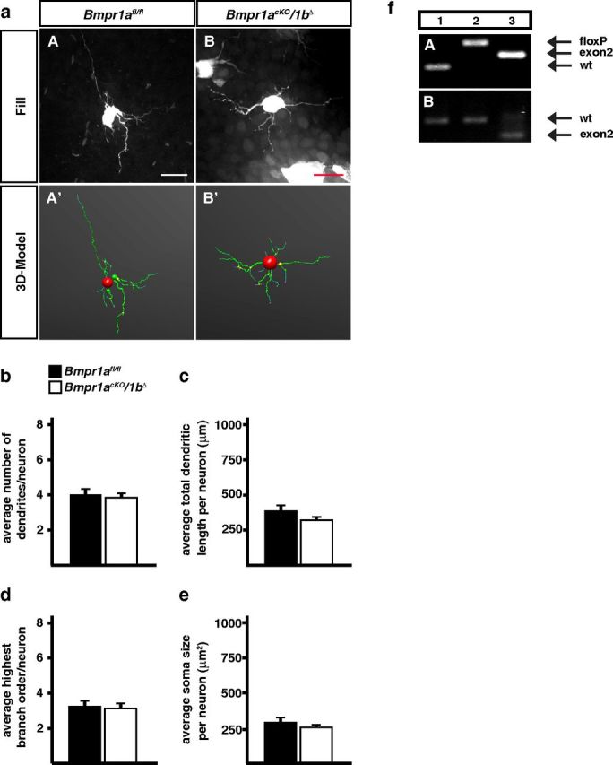
Dendrite morphology of P3 sympathetic neurons is not affected by Bmpr1 elimination in Bmpr1acKO/1bΔ mice. a, Sympathetic neurons of fixed SCGs were individually filled with fluorescent dyes (aA, aB) to obtain a 3D-model of the cells by confocal imaging and subsequent tracing (aA′, aB′). Quantification of morphological parameters showed no effects on primary dendrite numbers (b), total dendrite length (c), number of branch points (d), and soma size (e). Morphological analysis was performed on 31 control and 69 Bmpr1acKO/1bΔ cells. Error bars represent SEM. Statistical analysis by Mann–Whitney U test. Scale bar, 20 μm. f, PCR on genomic DNA from E14.5 stellate ganglia shows a virtually complete elimination of exon2 in Bmpr1acKO (fA, lane 3) as compared with wild-type (fA, lane 1) and Bmpr1afl/fl [fA, lane 2; Bmpr1a primers: Fx1 + Fx2 + Fx4, wt; 150 bp (Fx2/Fx4), floxed; 230 bp (Fx2/Fx4), Δexon2; 180 bp (Fx1/Fx4)]. Bmpr1a mRNA containing exon 2 (536 bp) is strongly reduced in E14.5 Bmpr1acKO stellate ganglia (fB, lane 3) as compared with wild-type or Bmpr1afl/fl ganglia (fB, lanes 1, 2) and Bmpr1a mRNA without exon 2 (372 bp) appears instead (fB, lane 3; Bmpr1a exon2 primers; Table 1).