
Figure 8.
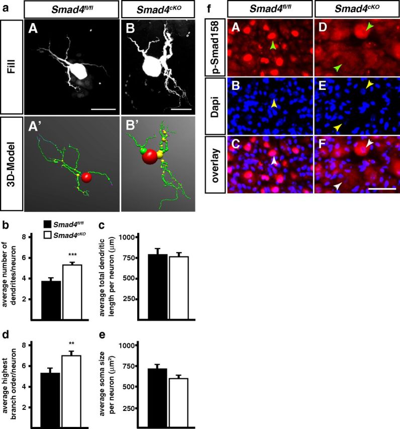
Dendrite morphology is altered in P90 Smad4cKO sympathetic neurons. a, Sympathetic neurons from Smad4fl/fl and Smad4cKO mice were injected with fluorescent dyes (aA, aB) to trace dendrite morphologies. b, d, The elimination of canonical TGF-β-signaling leads to a significant increase of primary dendrite numbers and highest branching orders in Smad4cKO neurons compared with the control (Smad4fl/fl). Total dendritic length (c) and soma size (e) are not affected in Smad4-deficient sympathetic neurons. Error bars represent SEM. Morphological analysis was done on 40 Smad4fl/fl and 38 Smad4cKO neurons and analyzed statistically by Mann–Whitney U Test; **p ≤ 0.01; ***p ≤ 0.001. Scale bar, 30 μm. f, The colocalization of pSmad1/5/8 and DAPI in Smad4fl/fl SCG neurons indicate a proper Smad4-mediated nuclear shuttling of activated R-Smads (fA–fC). In Smad4-deficient (Smad4cKO) mice phosphorylated Smads 1, 5, and 8 are predominantly located in the cytosol of sympathetic neurons (fD–fF). It is clearly visible that the nuclear area (fE; arrowheads) lacks pSmad1/5/8 immunoreactivity (fD, fF). Arrowheads indicate the position of the cell's nucleus. Scale bar, 20 μm.