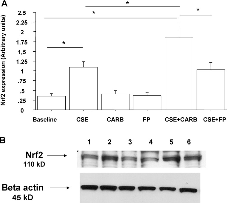
Fig. 8.
Effects of CARB and FP on Nrf2 nuclear expression in bronchial epithelial cells. 16-HBE (n = 5) cells were cultured in the presence and absence of CSE (10 %), CARB (10−4 M) and FP (10−8 M) for 18 h. Total proteins were extracted and nuclear proteins were collected and analysed for Nrf2 expression by western blot analysis. Membranes were then stripped and incubated with rabbit polyclonal anti-ß-actin. a Densitometric analysis of Nrf2 expression. Signals corresponding to Nrf2 on the various western blots were semi-quantified by densitometric scanning, normalised and expressed after correction with the density of the band obtained for β-actin. Data are expressed as arbitrary units ± SD. b Representative western blot performed on nuclear extracts. Lane 1, baseline; lane 2, CSE 10 %; lane 3, CARB (10−4 M); lane 4, FP (10−8 M); lane 5, CSE 10 % + CARB (10−4 M); lane 6, CSE 10 % + FP (10−8 M)

