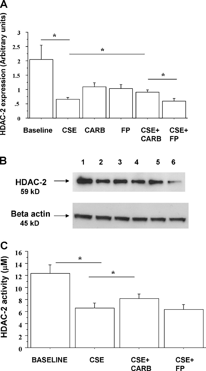
Fig. 9.
Effects of carbocysteine and FP on HDAC-2 nuclear expression in bronchial epithelial cells. 16-HBE (n = 6) cells were cultured in the presence and absence of CSE (10 %), CARB (10−4 M) and FP (10−8 M) for 18 h. Total proteins were extracted and nuclear proteins were collected and analysed for HDAC-2 expression by western blot analysis. Membranes were then stripped and incubated with rabbit polyclonal anti-ß-actin. a Densitometric analysis of HDAC-2 expression. Signals corresponding to HDAC-2 on the various western blots were semi-quantified by densitometric scanning, normalised and expressed after correction with the density of the band obtained for beta-actin. Data are expressed as arbitrary units ± SD. b Representative western blot performed on nuclear extracts. Lane 1, baseline; lane 2, CSE 10 %; lane 3, CARB (10−4 M); lane 4, FP (10−8 M); lane 5, CSE 10 % + CARB (10−4 M); lane 6, CSE 10 % + FP (10−8 M). c In some experiments (n = 3), nuclear proteins were immunoprecipitated and then assessed for HDAC activity. Data are expressed as micromolar ± SD using as reference a curve of the Fluor-de-Lys deacetylated standard (provided with the kit)

