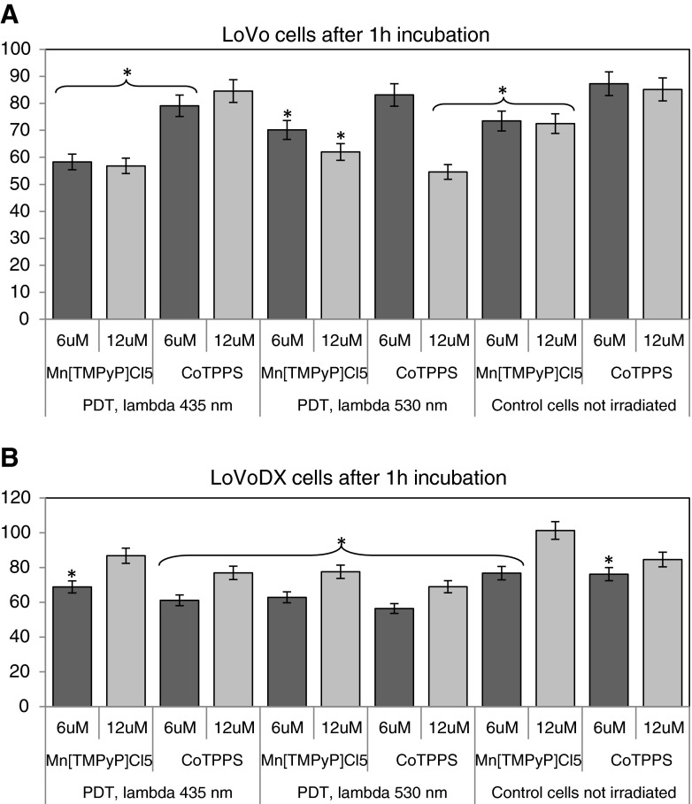
Fig. 3.
The photocytotoxicity assay performed in a LoVo and b LoVoDX cells. Cells were incubated with porphyrins during 1 h and then irradiated with two wavelengths (435 and 530 nm), then 24 h of incubation without the compound was followed. Each bar represents the mean of at least three separate experiments; bars are the standard errors, *p ≤ 0.005

