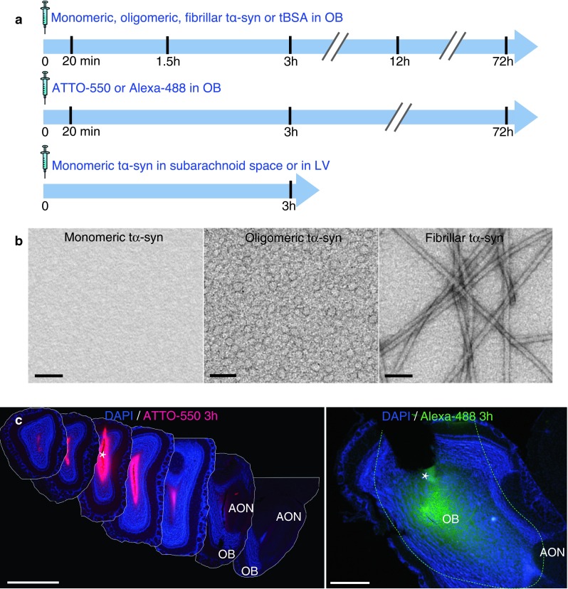
Fig. 1.

Experimental design and controls of diffusion. a Experimental design. In wild-type mice, we stereotactically injected α-synuclein (α-syn) into the olfactory bulb (OB), the lateral ventricle (LV) or at the level of subarachnoid space dorsal to the OB. Different molecular species of human recombinant α-syn tagged with ATTO-550 and S-tag (tα-syn) were injected: monomeric, oligomeric, or fibrillar tα-syn; or bovine serum albumin tagged with ATTO-550 (tBSA); or unbound ATTO-550 or Alexa-488 alone as control. We killed mice 20 min, 1.5, 3, 12 or 72 h after injection for histology. b Characterization of recombinant tα-syn. Negatively stained transmission electron micrographs of monomeric, oligomeric and fibrillar tα-syn. Oligomer samples are homogeneous, and do not contain any α-syn fibrils. Scale bar represent 100 nm. c Photographs of the injected region in the OB 3 h after injection of unbound ATTO-550 or Alexa-488 (dissolved in PBS) into the OB of mice. The images illustrate how far the injected solution diffused in the neural tissue. Left panel is a montage of coronal sections of a brain injected with unbound ATTO-550 (inter-section interval = 450 μm). Three hours after injection, ATTO-550 diffused from the injection site to lateral layers of the OB, and until the very anterior part of the AON, but not further. Scale bar 1 mm. Right panel is a low magnification picture of a sagittal section of a brain injected with Alexa-488. At 3 h after injection, Alexa-488 diffused from the injection site to the posterior part of the OB, the accessory olfactory bulb (AOB) and until the very anterior part of the anterior olfactory nucleus (AON), but not further. Dashed line represent the limit of Alexa-488 diffusion. Scale bar represent 500 μm. The white asterisk marks the injection site
