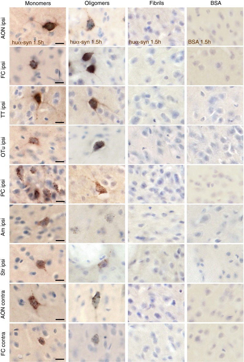
Fig. 4.

Transfer of tα-syn to other structures 1.5 h after injection into the OB. Images illustrating huα-syn or BSA staining at high magnification (scale bar 10 μm) in various brain areas. We detected huα-syn-positive cells in various brain structures 1.5 h after the injection of monomeric and oligomeric tα-syn into the OB. We observed huα-syn-positive cells in the ipsi- and contralateral anterior olfactory nucleus (ipsi/contra AON), in ipsi- and contralateral frontal cortex (ipsi/contra FC), in ipsilateral tenia tecta (ipsi TT), olfactory tubercle (ipsi Otu), piriform cortex (ipsi PC), amygdala (ipsi Am) and striatum (ipsi Str). On the contrary, we detected huα-syn-positive cells only in the ipsilateral OB when we injected fibrillar tα-syn. tBSA injected as a control protein into the OB, was not detected in the brain, except in the injected OB where we observed only a diffuse staining in extracellular space, but no obvious BSA-positive cell
