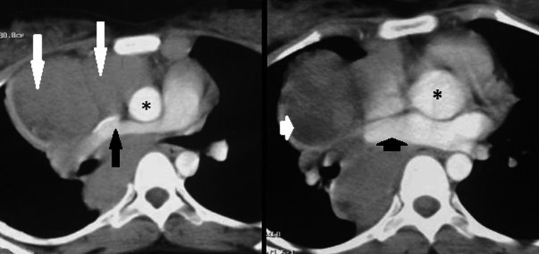
Figure 2.
Axial CT scans in a 21-year-old female with Hodgkin’s lymphoma show enlarged lymph nodes (long white arrow) compressing right pulmonary artery (long black arrow), SVC and right pulmonary vein (small black arrow). This compression effect is not seen in sarcoidosis. Asterisk represents ascending aorta. Also, note the hypodensity (small white arrow) in the central part of lymphadenopathy suggestive of cavitation.

