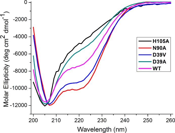
FIGURE 3.

Far-UV CD spectra of ISCU variants. All spectra were collected at 25 °C with solutions at pH 8. The CD spectra of ISCU(N90A) (red) and ISCU(D39V) (blue) indicate the presence of secondary structure, but the CD spectra of ISCU(H105A) (black) and ISCU(D39A) (green) suggest that little secondary structure is present. The CD spectrum of wild-type ISCU (magenta) is intermediate between those of the variants stabilizing the S- and D-states.
