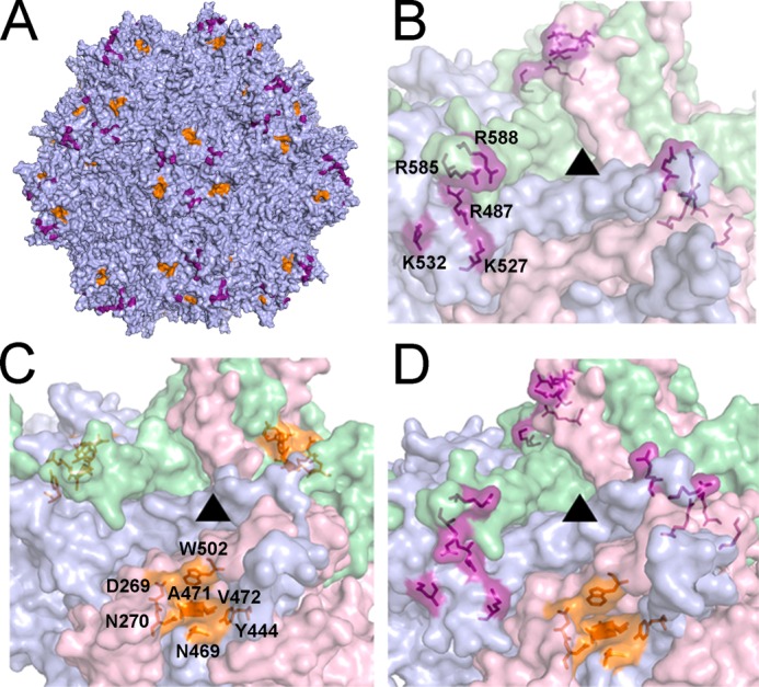
FIGURE 1.

Three-dimensional models of the dual glycan-binding AAV2G9 chimera and its parental strains AAV2 and AAV9. A, three-dimensional structural model of an intact AAV2G9 capsid with existing HS and “grafted” Gal binding residues shown in purple and orange, respectively. B–D, illustrations of the three-dimensional surface models of VP3 trimers at the 3-fold symmetry axes of AAV2 (B), AAV9 (C), and AAV2G9 (D) capsids. ▴ indicate the 3-fold axes of symmetry. Residues involved in HS binding (AAV2 VP1 numbering Arg-487, Lys-527, Lys-532, Arg-585, and Arg-588) and Gal binding (AAV9 VP1 numbering Asp-271, Asn-272, Tyr-446, Asn-470, Ala-472, Val-473, and Trp-503) are highlighted as in A.
