FIGURE 4.
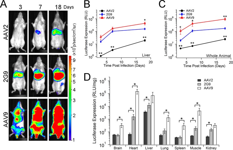
AAV2G9 mediates rapid onset and enhanced transgene expression in vivo. A, in vivo transgene expression kinetics of AAV2, AAV2G9, and AAV9 vectors packaging a CBA-luciferase transgene cassette. BALB/c mice (n = 4) were administered AAV vectors at a dose of 1 × 10¹¹ vg/animal through the tail vein, and bioluminescent images were collected at 3, 7, and 18 days post-injection using an Xenogen® Lumina imaging system. Representative live animal images are shown with bioluminescence on a rainbow-colored scale (1 × 10⁵-1 × 10⁶ photons/second/cm2/steradian). AAV2G9 maintains the hepatic tropism of AAV2 but demonstrates a more rapid and robust luciferase signal than both parental AAV strains. B and C, quantitation of the kinetics of light signal output (expressed as photons/second/cm2/steradian) was performed by marking regions of interest around images of the liver regions (B) and entire animals (C) obtained at different time intervals (n = 4). RLU, relative light unit. D, quantitation of luciferase transgene expression in major tissues from AAV2- (black), AAV2G9- (gray), or AAV9-treated (white) animals at 18 days post-administration (n = 4). BALB/c mice (n = 4) were administered AAV2, AAV2G9, or AAV9 packaging a CBA-Luciferase cassette at a dose of 1 × 10¹¹ vg/animal through the tail vein. At 18 days post-vector administration, animals were sacrificed to harvest brain, heart, liver, lung, spleen, skeletal muscle, and kidney tissues. Luciferase transgene expression in these tissue lysates was measured using a luminometer and normalized by the total amount of protein in each sample, determined using a Bradford assay. Statistical significance was assessed using one-tailed Student's t test. *, p < 0.05; **, p < 0.01.

