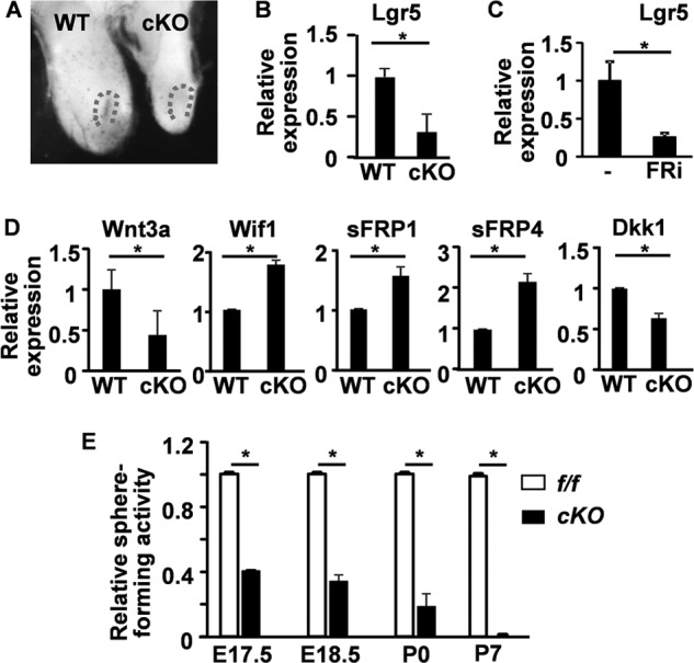
FIGURE 7.

Ablation of Fgfr2 suppresses Wnt signaling in the CLs. A, whole-mount LacZ staining showing Lgr5LacZ expression in the CL at postnatal day 0. Dotted lines indicate the CL. B–D, total RNAs extracted from the CL regions (B and D) at postnatal day 0 or DESC spheres (C) untreated or treated with 1 μm FGFR inhibitors were subjected to RT-PCR analyses of the indicated genes. E, sphere forming analyses of the CL cells harvested at the indicated stages. Data are means ± S.D. of triplicate samples. f/f, homozygous Fgfr2 floxed alleles; cKO, Fgfr2 conditional knock-out with Nkx3.1Cre; WT, wild type Fgfr2 alleles; E, embryonic; P, postnatal. *, p < 0.05.
