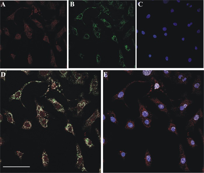
FIGURE 2.
Confocal evidence of mitochondrial and nuclear localization of FADS in primary cultures of neonatal rat ventricular cardiomyocytes. A, fixed and permeabilized neonatal rat cardiomyocytes were incubated with the polyclonal anti-FADS antiserum followed by incubation with an Alexa Fluor 568-conjugated anti-rabbit antibody (red). B, mitochondria were revealed with the monoclonal anti-Hsp60 antibody and an Alexa Fluor 488-conjugated anti-mouse antibody (green). C, nuclei were counterstained with Hoechst 33658 (blue). D and E, the colocalization of FADS with mitochondria (D) and nuclei (E) is depicted in white. Scale bar, 50 μm.

