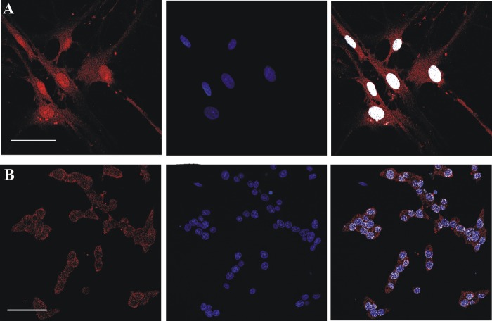
FIGURE 3.
Confocal evidence of a nuclear FADS localization in rat primary culture and cell lines. Fixed and permeabilized rat neonatal astrocytes (row A) and insulin secreting INS-1E β-cells (row B) were incubated with the polyclonal anti-FADS antiserum followed by incubation with an Alexa Fluor 568-conjugated anti-rabbit antibody (red). Nuclei were stained with Hoechst 33658 (blue). In the last panel of each row, the colocalization between FADS and the nuclear marker is depicted in white. Scale bar, 50 μm.

