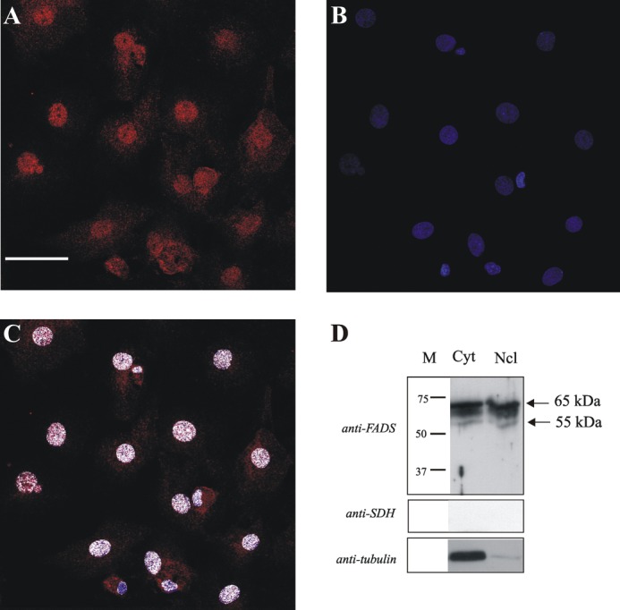
FIGURE 4.

Confocal microscopy and immunoblotting evidence of FADS localization in the nucleus of neonatal rat ventricular fibroblasts. A, fixed and permeabilized neonatal rat ventricular fibroblasts were incubated with the polyclonal anti-FADS antiserum followed by incubation with an Alexa Fluor 568-conjugated anti-rabbit antibody (red). B, nuclei were stained with Hoechst 33658 (blue). C, the colocalizion of FADS with Hoechst is depicted in white. Scale bar, 50 μm. Subcellular fractions of cardiac fibroblasts were obtained as described under “Experimental Procedures.” D, The cytosolic (Cyt) and nuclear (Ncl) fractions were analyzed by immunoblotting with the anti-FADS antiserum. After stripping, the same PVDF membrane was tested with anti-tubulin and anti-succinate dehydrogenase antibodies (anti-SDH), used as cytosolic and mitochondrial markers, respectively. The arrows indicate the position of the main anti-FADS-immunoreactive bands.
